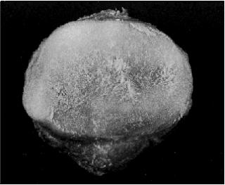
Slide 1shows the undersurface of a patella in a patient with chondromalacia patella. 320 x 263 pixels 13kb. This condition is most likely a precursor to OA and results in excess water in the cartilage (from Dieppe Textbook of Rheumatology).
Slide 2 "Ankle Bones 400 X 300 pixels 15kb Viewpoint model Worked in 3dStudio Max
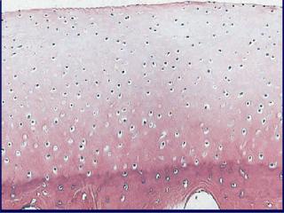
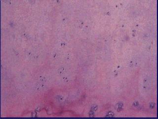
Slide 3: Shows abnormal cartilage in early OA where there has been a drop-out of chondrocytes secondary to necrosis (from Dieppe Textbook of Rheumatology).
The earliest changes in OA at the gross level are the irregularity of the cartilage surface, as shown here with the cartilage from the undersurface of the patella, followed by ulceration and then frank cartilage loss. However, as the disease progresses, all the joint structures are involved and results in “joint failure”.
Slide 4: Shows abnormal cartilage in which there are nests of chondrocytes forming in an attempt to repair the damage (from Dieppe Textbook of Rheumatology).
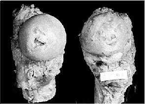
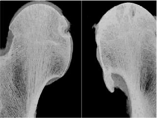
Slide 5:Shows two gross specimens of femoral heads (from Dieppe Textbook of Rheumatology).The hip on the left demonstrates mild OA with some focal loss of cartilage at the center. The hip on the right demonstrates severe OA with near complete loss of cartilage and the formation of large osteophytes.
Slide 6: Shows the MRI scans of the specimens in Slide 5(from Dieppe Textbook of Rheumatology)
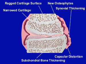
Several different pathophysiologic pathways can arrive at the structurally indistinguishable end result of joint failure .A normal synovial joint is "morphed " to show the characteristic changes of OA. There is loss of the cartilage thickness, as well as the formation of a ragged surface. New bone forms at the margins of the joint as osteophytes or bony spurs, while the subchondral bone thickens, and there is distortion of the capsule. Debris entering the joint space is taken up by phagocytes in the synovium, leading to synovial inflammation and thickening. Inflammation does occur in OA, but not to the extent seen typically with Rheumatoid Arthritis.
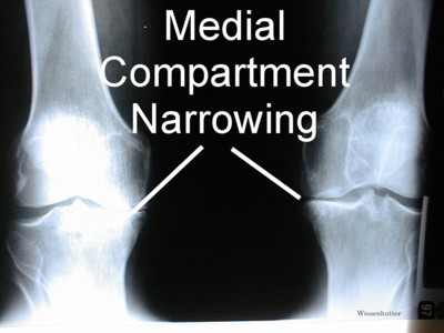
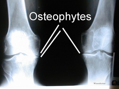
The characteristic changes seen with OA on a radiograph of the knees are shown. Note the prominent narrowing of the medial compartment and the prominent osteophytes.
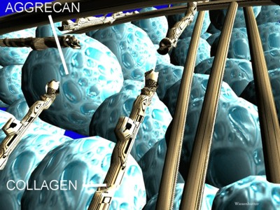
An early change in the structure of cartilage in OA is shown depicting damage to the collagen fibers. There is a loss of restraint on the aggrecan molecules and the molecules will expand, resulting in increased water content, or be lost from the matrix entirely. The end result is a decrease in the resilience of cartilage. Author's Note: The animations “Morphing of a Joint into Osteoarthritis”, and “Schematic Model of the Cartilage Defect in Osteoarthritis”, are two of my favorites. I made them both in 1998, and they were not difficult to construct. But I feel they hit home, and I will still visualize them today in my own mind when I'm discussing OA in the clinic, or when my own knee hurts.
Slide 7: "The characteristic changes that take place with an osteoarthritic joint are demonstrated on this drawing." 300 x 225 pixels jpeg 22kb drawn freehand with 3dStudio Max.